Back to: sex differences
Forward to: cyphopods
Gonopods
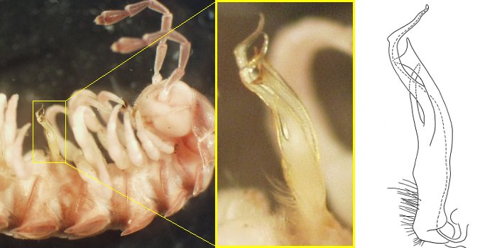
Tasmanodesmus hardyi (Dalodesmidae), Tasmania
Drawing shows right gonopod in medial view and dashed line marks prostatic groove (see below)
Drawing slightly modified from Mesibov (2004).
As shown on the body plans page, gonopods replace legpair 8 on ring 7 of adult males. The mating page describes how the gonopods are used to transfer sperm from the gonopores on male legpair 2 to the cyphopods on female ring 3.
Gonopods look nothing like legs (see images above), and they are by far the most variable anatomical structures in the Polydesmida. Their sizes, shapes, branching patterns and other details are extremely diverse, even within a single polydesmidan family. It would be silly for me to try to document that huge diversity here. Instead, I will describe and illustrate a few general principles of 'gonopodology'. For information about the gonopods of particular polydesmidan groups, go to the scientific literature on those groups.
One gonopod = one species
I am only half-joking when I write 'gonopodology'. Species of Polydesmida are recognised and diagnosed by their gonopods, and there are many scientific papers describing new species of Polydesmida in which the only illustrations are gonopod drawings.
Conversely, the only way to positively identify an adult male polydesmidan specimen is by comparing its gonopod with a description or drawing (see images above). If the specimen does not have gonopods (if it is a female or a juvenile, or a male with gonopods missing or damaged), it may not be identifiable to species, often not to genus, and sometimes not even to family.
The good news is that gonopods differ so much between species, and so little within species, that they are almost like fingerprints: when you find a match, the match will usually be a good one (see images below). If you cannot find a good match, you may have a new species.
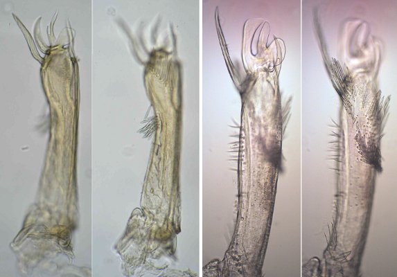
In 2008, Jörg Spelda (Zoologische Staatssammlung München, Germany) sent me two images (above left) of a gonopod. The millipede they came from had been collected years before by Martin Baehr during a visit to Tasmania. I was able to identify the millipede as Tasmaniosoma compitale (Dalodesmidae) just by its distinctive gonopod (images above right, from a specimen in the Queen Victoria Museum and Art Gallery in Tasmania), although it was not until 2010 that the species was formally named and described. [The two gonopods have been imaged at different angles, and the minor differences between the two are within the natural range of variation in this species.]
There are a few exceptions to 'one gonopod = one species'. These are species which have very similar gonopods but which clearly and consistently differ in 'non-gonopodal' (or somatic) anatomy, and which overlap in their geographic ranges.
Constant elements
Although polydesmidan gonopods can differ enormously in form and detail, they almost all have the same basic construction*. To begin with, the two gonopods sit in an aperture on ring 7. The shape of the aperture varies, as does its size. The aperture in Tasmanopeltis grandis (image below, left) is ovoid in ventral view and fairly small: about one-third the width of the ring 7 prozonite. Parts of the aperture margin may also project ventrally. In other words, on an upside-down male the margin will look 'raised' (image below, top right). A peculiarity of Paradoxosomatidae is that the aperture is constricted in the middle by projections from the aperture margins (image below, bottom right).
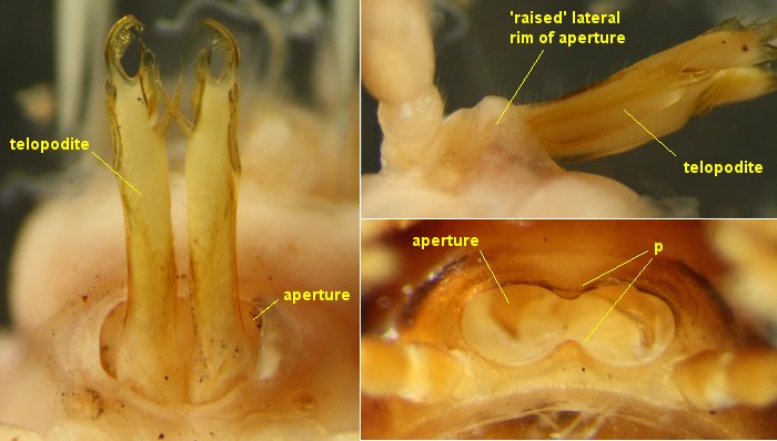
Gonopods of Tasmanopeltis grandis (Dalodesmidae), Tasmania in posteroventral (left) and left lateral (top right) views. The gonocoxae are entirely contained within the aperture and are not visible
Posteroventral view of aperture of Somethus tasmani (Paradoxosomatidae), Tasmania (bottom right) with gonopods removed, showing aperture rim projections (p). See below for lateral view with gonopods in place
The gonopod itself usually consists of a gonocoxa and a telopodite. In some Polydesmida the telopodite pivots on the gonocoxa, while in other Polydesmida the two elements are fused together. The gonocoxae may be small or massive, 'hairy' with setae or relatively bare, entirely within the aperture (image above, left) or largely outside the aperture (image below, left), attached to the aperture rim or free. In some polydesmidan groups the two gonocoxae are fused together into a syncoxite. In other groups the two gonocoxae are only lightly joined along their median edges, and in Paradoxosomatidae the two gonocoxae are entirely separate.
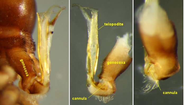
Somethus tasmani (Paradoxosomatidae), Tasmania. Right lateral view of gonopods in place (left),
and medial (centre) and posterior (right) views of detached right gonopod
Telopodite features
Most polydesmidan males use their gonopod telopodites to pick up sperm-containing fluid from the gonopore on ring 2. The gonocoxa is connected to the telopodite through a short tube called the cannula (image above, centre and right), which empties into a narrow prostatic groove on the telopodite. Just how the gonocoxa, prostatic groove and picked-up sperm work together is not known. The prostatic groove is often like a tunnel, because the two rims of the groove may touch each other at the surface of the telopodite, enclosing a cavity. For this reason it is usually easy to trace the course of the groove in a telopodite which has been cleared of soft tissue and thus made transparent, and often hard to trace the groove in a scanning electron micrograph, which only shows the telopodite surface.
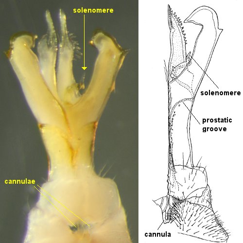
Posterior views of gonopod of Gasterogramma psi (Dalodesmidae), Tasmania. The cannula enters the base of the telopodite on the telopodite's medial side, and the base of the small, worm-like solenomere is also close to the medial side. The prostatic groove, however, does not join the two in a straight line. Instead it swings laterally on the posterior surface of the telopodite and circles around the base of the large lateral process on the telopodite before reaching the solenomere. The prostatic groove often takes an unexpectedly indirect path on the telopodite in Polydesmida. Drawing at right modified from the original by the late C.A.W. Jeekel (1982).
When the prostatic groove ends on a particular branch of the telopodite, that branch is called a solenomere (solenomerite in the older Polydesmida literature). Many different names are in use by Polydesmida specialists for other parts of the telopodite, including podomere names such as 'tibiotarsus'. There is no Polydesmida-wide system for naming telopodite parts, and best practice at the moment is to follow the naming system used by Polydesmida specialists who have previously worked on the group in question. For instance, in some polydesmidan families the basal portion of the telopodite has setae and is slightly separated from the distal part of the telopodite by a constriction. This basal portion is usually called the prefemur of the telopodite, and the whole of the telopodite distal to this prefemoral portion is called the acropodite. For a clearly illustrated example of this kind of gonopod, see Paul Marek's webpage on Brachoria (Xystodesmidae).
There are some Polydesmida with a prostatic groove but no cannula (Rhachodesmidae) and others with neither a cannula nor a groove (the genus Agathodesmus in Haplodesmidae, and Tridontomus in Tridontomidae). It would be interesting to study mating in these groups! Another exceptional group is the family Holistophallidae, in which the gonopod is in one piece.
Drawing gonopods
Telopodite branches often have complex, curving shapes which cannot be properly represented in a single, two-dimensional view. Furthermore, some telopodite parts may be partly hidden from view by other parts. A good way to show the topology of a complicated telopodite is with a series of drawings from different viewpoints (see example below). To make such drawings I use a microscope with a square grid (reticule) in the eyepiece. I transfer what I see in each square to the corresponding square on a piece of paper marked with a square grid. A camera lucida attachment for a microscope can also be used, but is much more expensive.
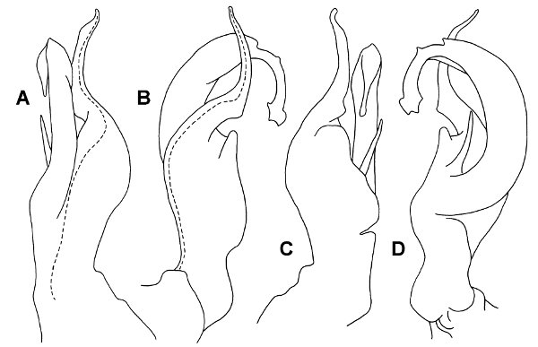
Gephyrodesmus coolahensis (Dalodesmidae), New South Wales, Australia
Right gonopod telopodite in anterior (A), lateral (B), posterior (C) and medial (D) views
Dashed line marks course of prostatic groove; setae not shown
Drawings from Mesibov (2008)
When making gonopod drawings, be sure to include the path taken through the telopodite by the prostatic groove. This path varies between genera and species in ways that are very useful for classifying and identifying.
*The strange cave-dwelling Dobrodesmus mirabilis, from Brazil, has very differently constructed gonopods, with a flagellum arising from the coxa, no cannula and no prostatic groove (Shear et al. 2016).
Jeekel, C.A.W. 1982. Millipedes from Australia, 4: A new genus and species of the family Dalodesmidae from Australia (Diplopoda, Polydesmida). Bulletin Zoölogisch Museum, Universiteit van Amsterdam 9(2): 9-15.
Mesibov, R. 2004. Redescription of Tasmanodesmus hardyi Chamberlin, 1920 (Diplopoda: Polydesmida: Dalodesmidae). Myriapodologica 8(3): 21-36.
Mesibov, R. 2008. The millipede genera Gephyrodesmus Jeekel, 1983 and Orthorhachis Jeekel, 1985 in southeastern Australia, a new Lissodesmus Chamberlin, 1920 from Victoria, and observations on male leg setae, spinnerets and metatergite sculpture (Diplopoda: Polydesmida: Dalodesmidae). Zootaxa 1790: 1-52.
Shear, W.A., Ferreira, R.L., Iniesta, L.F.M. and Marek, P. 2016. A millipede missing link: Dobrodesmidae, a remarkable new polydesmidan millipede family from Brazil with supernumerary rings (Diplopoda, Polydesmida), and the establishment of a new suborder Dobrodesmidea. Zootaxa 4178(3): 371-390.
Back to: sex differences
Forward to: cyphopods